We listen to the heartbeat of the child during pregnancy
Procedures that allow you to hear the heartset of the fetus: ultrasound, echocardiography, auscultation and ktg. The rate of heart rate at different terms: what is evidenced by their participation and gentle
Fetal heartbeat – the main indicator of its viability. Any rhythm changes indicate the emergence of adverse factors. Therefore, doctors control this process throughout the entire pregnancy and during childbirth.
Heart features from the fetus
 Approximately 4 weeks are laid by the heart of the heart, which is a hollow tube, but after 7 days the first ripples appear. By 9 week, the body becomes four-chamber. However, in the womb, the baby does not breathe himself, but receives oxygen from the mother, so his heart has some features, for example, a hole between the atrialists and the arterial duct, which are closed after birth.
How to hear heartbeat crumbs?
Listen to how the heart beats your baby, you can in several ways:
- Ultrasound (ultrasound research);
- Echocardiography (echocardiography);
- Auscultation (listening);
- KTG (cardiotokography).
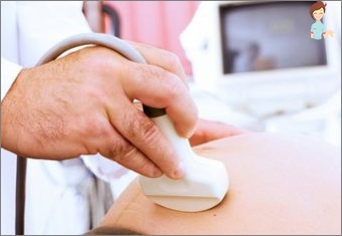
Ultrasound
In the first trimester, the fetal heart rate is determined by weeks using ultrasound research. Normally, during a transvaginal ultrasound, the reduction of the heart is detected by 5-6 weeks, and during transabdominal – from 6-7.
The heart rate in the first trimester depends on the timing:
- Up to 8 – from 110 to 130 shots per minute;
- Fetal heartbeat in 9-12 weeks – 170-190;
- From 13 to childbirth – 140-160.
Frequency changes are related to the formation of the function of the nervous system, or rather, the part responsible for the operation of the organs. An adverse signs are the impact of frequency up to 85-100 beats per minute, as well as significant increase (up to 200).
In such cases, the reason determined and eliminates the impaired cardiac rhythm. If the embryo reached 8 mm, but there are no heart abbreviations, it testifies to undeveloped pregnancy. In order to confirm or disprove the diagnosis, re-ultrasound after 5-7 days.
In the second and third trimester, during the passage of planned ultrasound studies, it is determined:
- Heart location. Normally, it is placed on the left and takes about a third of the sternum during transverse scanning;
- Frequency (heart rate of the fetus – 140-160);
- Character of abbreviations (rhythmic / arrhythmical).
The frequency of abbreviations in late terms largely depends on foreign factors, for example, the movements of the fetus, physical exertion of the mother, the impact on pregnant variety of factors (heat, cold, diseases).
If the fruit does not receive enough oxygen, then at the beginning of the heart rate increases over the norm (tachycardia), and then, after the deterioration of the child’s condition, falls below 120 (bradycardia).
To reveal the vices of the heart, use the so-called «four-chamber slice» – ultrasound, allowing you to see all 4 organs of the organ. With this method, about 75% of pathologies are found. If there is a need for additional studies, then prescribed the echocardiography of the fetus.
Echocardiography
 This procedure is a special type of ultrasound research. EchoCG – a comprehensive way that allows you to fully explore the heart. It includes in addition to the standard two-dimensional ultrasound and other scanner operation modes: M-mode (one-dimensional) and dopar mode (for the blood flow in different departments). EchoCG allows you to explore the structure of the heart and blood vessels, as well as their functions.
This event is carried out according to the testimony:
- Sugar diabetes in the future mother;
- Infections transferred during the battery period;
- Age of pregnant more than 38 years;
- Congenital heart defects (UPU) in mothers;
- Child growth delay;
- In the history of the birth of children with UPU;
- Changes in the heart, discovered ultrasound (disturbed rhythm, increase in size and pr.);
- Other pathologies, including genetic diseases that are often combined with heart defects.
Optimal deadlines for EHOCG – 18-28 weeks. In the future, the imaging of the heart is difficult, as the number of accumulating waters is reduced, and the child increases in size.
Auscultation
This method is the easiest. This requires an uncomplicated device for Listening to the palpitations of the Future – Obstetric Stethoscope. From the usual it is distinguished by a wide funnel, which is applied to the belly of the future mother, and on the other hand, listen.
Since the invention of the stethoscope, its form has almost changed. Standard device is made of wood, but now there are plastic and aluminum products.
Cardiac tones start to listen about from 18 weeks. As the child develops in the womb, they are heard more and more. On each planned examination, when the fetal appears with a stethoscope of heartbeat, the doctor necessarily conducts listening, and also this phenomenon is controlled by an obstetrician during childbirth.
With auscultation, other sounds are tested:
- Intestinal noises (bubbles, iridescent, irregular);
- Reduction of uterine vessels, aorta (coincide with a woman’s pulse);
- The point of the best listening to heart rate, the nature and rhythm of the heart abbreviations is determined;
- With a head preview, the tone is clearly audible below the navel, with a transverse one level with it, when pelvic – above;
- Lisviar rhythmism. Arrhythmical are characteristic of heart defects and oxygen deficiency (hypoxia);
- Tones may not listen because of many- or lowland, obesity, multipleness and increased fetal activity.
In the process of childbirth, the obster conducts listening to the fetal heartbeat every 15-20 minutes, at the same time assessing the rhythm before and after the fight to determine how the fruit reacts to it. The doctor also listens to the heart rate after each sweat, since at this time the muscles of the uterus, the pelvic bottom and the abdominal wall are reduced by the woman in labor, which leads to a decrease in oxygen access to the fetus.
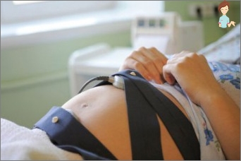
Cardiography
 The method allows you to objectively explore the heart of the child after 32 weeks of pregnancy. Cardiography registers not only heartbeat, but also cutting uterus. In modern devices there is a function of recording the motor activity of the kid in the womb.
When conducting a procedure, a woman should lie on the back, side or sit. The procedure is carried out both before delivery and during such. The sensor is fixed in the place of best listening to the tones and leave for 1 hour. The results allow the doctor to evaluate the heart rate, its change in response to the fight and movement of the child.
The need for CTG appears in the following situations:
- Fever in a pregnant woman with a temperature above 38;
- Severe gestosis;
- Scar in the uterus;
- Chronic diseases (hypertension, diabetes);
- Calling of childbirth (induction) / relaxation with weak generic activities;
- Low or multi-way;
- Premature aging placenta;
- Intrauterine delay in development;
- Change in the frequency and character of heartbeat with auscultation;
- Violation of arterial blood flow.
After the CTG, the doctor assesses: the average heart rate (norm – 120-160), rhythm variability (permissible oscillations – 5-25 beats per minute), change in frequency due to bouts or fetal movements,.
The increase in the heart rate in response to the contraction of the uterus is a positive sign. Defense testifies to hypoxia, fruit-placental insufficiency, but normally occurs in the pelvic position of the child.
The heartset of the fetus in the late periods of weeks is not determined, but if necessary, the CGT can be conducted repeatedly.
Studies using a variety of methods can be carried out throughout the period of pregnancy, as well as during the generic process, and allow you to assess the condition of the baby, to begin treatment in a timely manner in case of detection of pathology and resolve the issue of the delivery.
How to hear the heartset of the fetus while being at home
 For listening, you can use the above-signed stethoscope, but someone else can take advantage of it, and not the most pregnant. For future mothers, it seems possible to hear the heart rhythm of the baby using special devices designed for use at home, such as autonomous fetal dopplers (monitors).
Such a device can take advantage of both the woman and other family members. It is quite safe for the health of the future mother and child, allows you to hear a heartbeat from about 12 weeks of pregnancy without leaving home, at any convenient moment.
Enjoy the sweet sounds of the heart of your crook! Quick and easy to give birth!


