Screening during pregnancy
How the first, second and third screenings are held. What are screenings and how to decipher them?
Many women very word «screening» inspires fear. In fact, there is nothing terrible, dangerous or incomprehensible. This word comes from the concept «screening». That is, in medicine, the screening is understood as a mass examination in order to identify pathology as mom and a child. That is, screening during pregnancy is a survey of future mothers, to determine the health and full development of their future kids.
With this study in the early and middle stages of pregnancy, the so-called congenital pathologies identify. That is, you will know in advance whether your baby or he will need help doctors after birth.
 The main observations are aimed at identifying such complex pathologies as a Down syndrome (trisomy 21), Edwards syndrome (trisomy 18) and defects of the nervous tube (DNT).
In addition, the prenatal screening allows you to learn about such pathologies in advance of such pathologies as Cornel de Lange syndrome, Smith-Lemlice syndrome, non-polar triploidy, Patau syndrome.
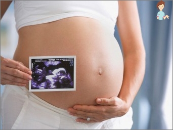
Screening during pregnancy consists of a number of analyzes and is carried out in parallel with the ultrasound study of the kid with additional measurements.
Consider everything in order and Read more.
First screening
The first comprehensive screening during pregnancy includes biochemical analysis of venous blood for determining the content ?-Subunits of human chorionic gonadotropin and rarr-a – plasma protein associated with pregnancy.
This analysis is done in order to identify in the early stages of deviations from the norm in the development of the embryo. If analyzes showed no norm, it can talk about congenital pathology of development or genetic anomaly in a child.
In this case, the mammy is directed to additional studies – such as amniocentesis (spindle water by a special needle take on the analysis) and biopsy of the Vorsin Chorion.
At the first screening, as a rule, women are sent from a risk group:
- Over the age of 35;
- Those in which there are Down syndrome or other genetic pathologies;
- Those in the past were miscarriage, stillbirth, frozen pregnancies, children with congenital pathologies or genetic anomalies;
- Women who at the beginning of pregnancy suffered viral diseases, took the drugs dangerous for embryos or exposed to radiation.
On the ultrasound of the first screening (11-13 week of pregnancy) are determined by:
- The amount of embryos in the uterus in a woman, and their ability to survive;
- The term of pregnancy is specified;
- The coarse malformations of the development of the organs in the child are revealed;
- The thickness of the collar zone is determined, which implies the measurement of subcutaneous fluid in the rear region of the neck and shoulders;
- The nasal bone is investigated.
The deviation from the norm at the first screening indicates the likelihood of the risk of various chromosomal and some non-chromosomal deviations. It is worth noting that the increased risk is not a reason for excitement about the defects in the development of the fetus.
These studies suggest a narrower monitoring of pregnancy and full-fledged child development. Often, if the result of the first screening showed an increased risk for some indicators, then the conclusions can be done only after passing the second analysis. And with serious deviations from the norm, a woman consults a geneticist.
Second screening
The second comprehensive screening during pregnancy is performed from 18 to 21 week. This study implies «triple» or «Four test». It is performed in the same way as in the first trimester – a woman gives a biochemical analysis of venous blood.
But, the second screening involves the use of analysis results to determine three or less than four indicators:
- Free beta subunit hgch;
- Alpha fetoprotein;
- Free estriol;
- Four test – Ingibina A.
 The interpretation of the results is based on the deviation of the obtained indicators from the average norm according to certain criteria. Calculations are carried out by a computer program, then analyzed by a doctor, which takes into account the set of individual parameters (the race to which the pregnant and father of the child owns; the presence of chronic, including inflammatory diseases; amount of embryos; Women’s body mass, presence or absence of bad habits).
Similar secondary factors may affect the change in the values of the test indicators.
For maximum reliability, the results are necessarily compared with the data of prenatal screening in the first trimester. And in addition to biochemical analysis, an ultrasound is also held on which the already formed kids are watching.
If, according to the results of the first and second screenings, deviations are revealed in the development of the baby, the woman can offer repeated research or guide to a consultation to a genetics specialist.
Third screening
Third screening is carried out at 30-34 weeks of pregnancy. It is held in order to assess the functional state of the baby, and make sure that the child and the placenta interact effectively.
Also estimated the position of the kid. This will depend on how he will behave in childbirth, and what tactics of childbirth to choose the doctor. In addition, the condition and location of the placenta is estimated, which is also important for future labor.
During the third study, the woman again prescribed a triple biochemical analysis of venous blood, and can also be submitted to additional research, such as Dopplerography and Cardiotocography.
Dopplerograph allows the doctor to estimate the blood flow power in the uterine and placental vessels, as well as in the trunk vessels in the child. This gives an understanding of how much oxygen and nutrition gets a baby and whether they grabs them.
Cardiotokography (CTG) registers the frequency of heart cuts in a child, even in the process of childbirth. Child’s pulse is determined by a special sensor. This test is carried out only after the 32nd week, because in terms of the wound 32 weeks the results will be unreliable.
The first, second and third screenings are held in order to assess the health of your baby and his readiness for childbirth, because it should be actively «work». Studies allow you to prevent some possible problems.
After all, any problem is easier to prevent than then long and persistently treat. In any case, to comply with the prescribed surveys during the same time of pregnancy. Objectively, this is the only opportunity to learn about the health of the kid in advance.
And do not believe «People» rumors that ultrasound is harmful to kid or annoys it. This is an absolute not true, especially if you are examined in a clinic that has modern equipment and high-class specialists.

Even if the doctors said that you have a high risk of pathologies – do not despair. It is not always something purely serious, requiring global solutions to maintain or no pregnancy.
Sometimes a child can find pathologies requiring surgical intervention after birth.
And, if you and the doctor know about it in advance, then the chances of the most effective treatment and excellent health of your baby increase significantly.
You, as a mother, are primarily responsible for the future of your baby, so take off the screenings with full responsibility. Find a specialist who competently and available to explain to you what is happening with you and your chance and thereby dispel all your fears and doubts.
And let everything and your baby be fine!


