All about hip dysplasia in newborns
Why a newborn baby displays of hip joints – features of the structure and causes. How to determine and cure a disease – symptoms, signs, as well as treatment of dysplasia in children

With dysplasia (congenital dislocation of hips) in newborns, parents face quite often. The disease is characterized by underdevelopment or improper formation of joints.
If the baby put such a diagnosis, you need to immediately begin treatment so that there are no disorders in the work of the musculoskeletal system.
Features of the structure of hip joints
Sustaines in a child even with normal development differ from the anatomical parameters of adults, although, in both cases, the joints serve as a connecting link between the bones of the hip and the pelvis.
The upper part of the femoral bone has a spherical head on the end, which is included in a special recess in the pelvic bone (accommodation depression). Both structural parts of the joint are covered with a cartilage cloth preventing bone wear, which contributes to their smooth slip and depreciation acting on the joint.
Task joints – Provide body turns in different directions, flexion and extension of the limbs, hip movement in space.

The godfather of the hip joint in children is not in the inclined position, as in the organism of an adult, but Almost vertical and has a more flat configuration. The head of the bone is held in the cavity through bundles, the master’s lips and the joint capsule, which worst almost completely the neck of the thigh.
Children’s bundles have significantly greater elasticity, than in adults, and most of the hip area consists of cartilage tissue.
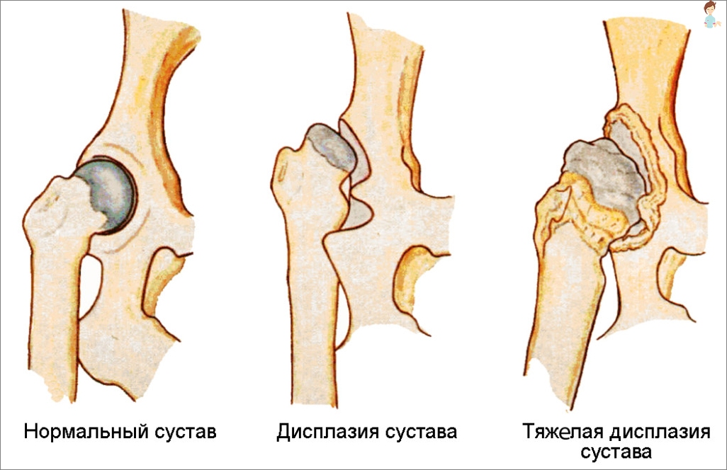
Dysplasia of joints in children is classified by experts in terms of the development of the joint development of the standard parameters
| The immaturity of hip
Sustav |
The immaturity of the children’s joint is not yet pathology, because in the future its development can reach the norm. Detect immaturity, you can only with the help of an ultrasound that shows a slight compaction of the masterpiece. |
| Present | Is the initial stage of dysplasia. Manifests itself with a small pathology in the joint connection, but the malformation is not observed. |
| Subways | Characterized by shift head bone. Because of this, it is only partially in the depression, which also has a form defect. |
| Dislocation | Hip head is outside the depression. |
Causes of displays of hip joints in children
There are several factors that are in one way or another affect the formation of dysplasia in a newborn:
- Hereditary factors, When pathology arises due to abnormalities in the body under the influence of genes. That is, the disease is laid at the embryo level and prevents the normal development of the fetus.
- Restriction of the free motion of the fetus in the womb, caused by the wrong position of the child in the uterus of the uterus (Malovodie, Multiple pregnancy and T.D.).
- Up to 50% of dysplasia occurs due to the large size of the fetus, As a result, it shifts from a normal anatomical position (Pelvic preview).
- Paul baby. Most often the disease is found in girls.
Often the cause of dysplasia are factors whose carriers are the following mother:
- Infectious or viral infections that were overcome pregnant woman.
- Unbalanced nutrition, lack of group vitamins B and D, as well as calcium, iodine, phosphorus and iron.
- Violation of metabolism in the body.
- Toxicosis at the early or late stage of pregnancy.
- Incorrect lifestyle of the future mother (smoking, alcohol).
- Problems with cardiovascular system.
Important! Inexperienced parents often accuse the doctors who give birth in the fact that they are due to non-professional actions allowed displays. In fact, the pathology of the hip area develops During the growth of the fetus in the womb, and not in the process of childbirth.
How diagnose displasion of hip joints in children – symptoms and signs of the disease
If the pathology in the hip joint is pronounced quite brightly, the diagnosis of the baby is put in the maternity hospital.
Unfortunately, to identify the ailment in the first days after birth can not always. Cross defect in the joint does not cause any inconvenience, so he behaves calmly, and to suspect the disease on the behavior of the child, parents cannot.
Signs of illness reveals a doctor when conducting a medical examination. In addition, for some clear indicators, a mother can determine the pathology independently.
The presence of the disease indicate such signs as:
| Asymmetry of inguinal or buttocks | If you put a baby on your back or tummy, folding on the legs are asymmetrically, and on one foot there may be more than on another |
| Symptom click | A characteristic clock when breeding legs to the side occurs even with a small hill of the joint. This is a clear sign of pathology, but after 7-10 days after birth, click disappears. |
| Limited breeding hips | In a healthy newborn baby, the legs bent in the knees are bred to the sides, forming the angle between the hips 160-170O. In a child with a dysplasia, a leg with the affected joint is not fully given. |
| One leg is shorter than another | With the pathology of the hip joint of the child’s leg in an extended position have a different length. |
Important! Sometimes there may be cases of asymptomatic course of the disease. To not start the process, visit orthopedes. In case of doubt, the doctor prescribes an ultrasound study or radiography.
If you do not detect pathology in the early stages on time, the hip head will shift until dislocation is formed, and the change in the musculoskeletal functions of the joint will begin.
Features of the treatment of hip dysplasia in children
To the treatment of dysplasia, you need to start immediately after diagnosing. The main task of eliminating pathology is to ensure that the head of the bone of the hip is correctly located and fixed in the masterpiece.
For this, such treatment methods are used as:
| Massage treatments | In order not to harm the child for massage, you should contact an experienced specialist. The joints and bones of the newborn are very podiatiles, any incorrect impact on them can lead to a violation of the normal functioning of the musculoskeletal system.
When using a massage, you need to systematically monitor the process, conducting an ultrasound through a certain number of sessions. The frequency of checks establishes a doctor. Ultrasound gives an objective assessment of the treatment process and, in the event of the ineffective method, other procedures are immediately appointed. |
| Wide swab | The wide swarer method helps the normal development of hip joints, prevents the appearance of a subluxation and dislocation of the thigh head, reduces the risk of operation.
Widespread swords of the child’s feet fixes them in a slightly bent position, and the hips are bred under the desired angle. With a wide swellant use method 3 diapers. One of them is folded in several layers so that its width is 20 cm. and pave between the legs of the child. So they are bred in different directions. The second diaper is a triangle, one angle is laid between the legs, and the two remaining wipes the feet of the child, bringing them out 90O. In 3, the diaper wrap the kid on the belt, while the legs slightly pull up so that the faces of the crumbs are not connected. Such swaddling allows the child to feel comfortable. |
| Use of orthopedic devices |
|
| Medical exercises | LFK strengthens the kid’s muscles. Exercises are performed by putting the child on the back:
Each exercise makes 8-10 times. |
In addition, the attending physician may prescribe paraffin wraps and electrophoresis with calcium and phosphorus to strengthen the joints.
If at least the slightest suspicion of pathology arose, you need to urgently contact the specialists and begin treatment!
Lady-Magazine site.COM warns: information is provided solely for informational purposes, and is not a medical recommendation. In no case do not self-medicate! If you experience health problems, consult your doctor!


